-

FOR2848 in a nutshell
Overview of the projects and methods used in this research unit. -

Live-Cell staining of Cristae
Live-Cell STED image of the inner mitochondrial membrane (Stephan et al., 2019, Scientific Reports) -
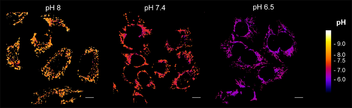
Ratiometric imaging of mitochondrial pH
Matrix pH measurement with the pH sensor mt-sEcGFP in HeLa cells. (Rieger et al., 2021, EMBO Reports) -
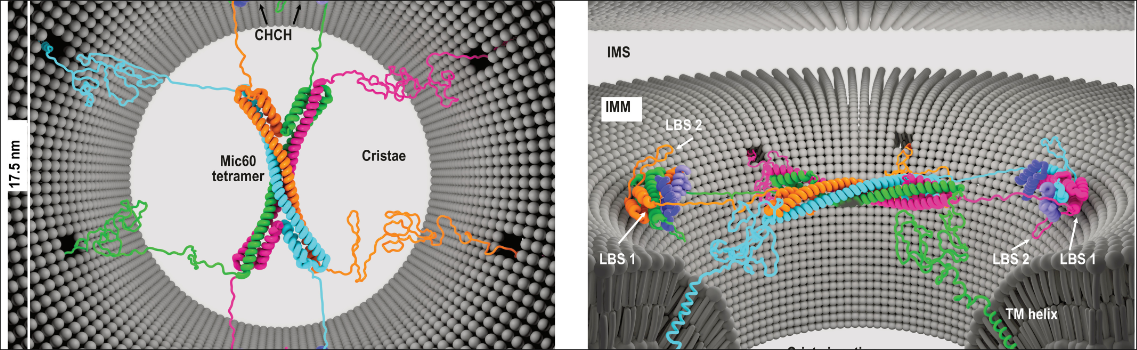
Model of Mic60-Mic19 function at crista junctions
Top view and side view showing the proposed architecture of the Mic60-Mic19 complex at crista junctions. (Bock-Bierbaum et al., 2022, Science Advances) -
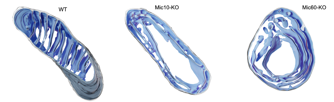
3D reconstruction of mitochondrial inner architecture
Reconstruction of electron tomography data showing altered crista morphology in Mic10- and Mic60-knockout cells. (Stephan et al., 2020, EMBO Journal)
Research Unit 2848
The inner membrane of mitochondria exhibits a unique three dimensional structure. It is also exceptionally rich in proteins, most of which facilitate transport and catalytic processes. Currently, it is assumed that the structure of the inner membrane enables dedicated protein and lipid environments resulting in the spatial organization of protein functions. Accordingly, protein function and membrane topology appear to be tightly intertwined with each other in mitochondria. Considering this, it is surprising that we still lack detailed insight into how the inner membrane is shaped. Moreover, information on the actual distribution of protein functions in the inner membrane is very scarce at the best. To this end, experimental approaches that provide temporal and spatial information of inner membrane shape and protein localisations are essential to assess and eventually understand how membrane shape and functional organization are brought about. In this research initiative, we take a decisive step to tackle fundamental questions on mitochondrial ultrastructure and bring together researchers with unique expertise in mitochondrial imaging technologies with scientists addressing central biological questions on mitochondrial functions. We expect that the combination of functional analyses with state-of-the-art microscopy will allow us to obtain a detailed view on the organization of the inner membrane, to address the question on how proteins support the shaping of the inner membrane, and to define how these processes are interconnected and regulated in the cellular context.